产品展示更多>>
- Rabbit anti-IL-1β Polyclonal Antibody

- Rabbit anti-IL-1β Polyclonal Antibody
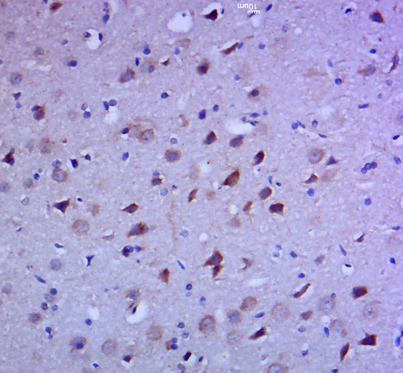
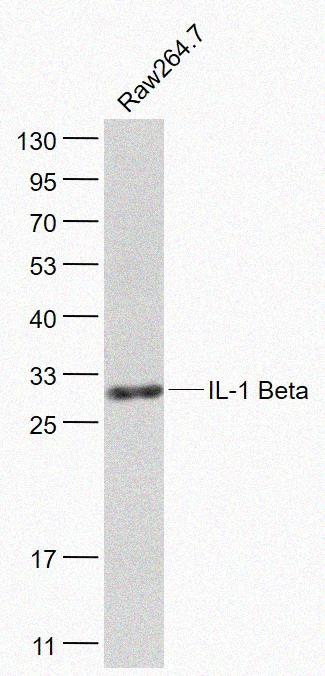
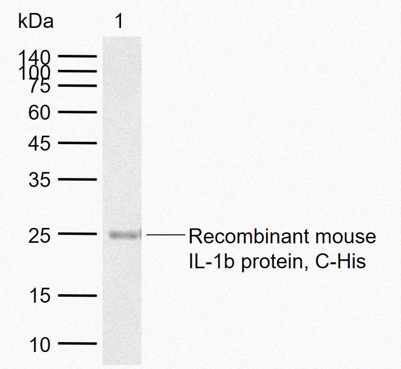
概述 别名 IL-1 β抗体;Catabolin; H1; IL-1β; Hematopoietin 1; IFN beta inducing factor; IL 1; IL 1 beta; IL 1B; IL1B; IL1F2; Interleukin 1 beta; Interleukin 1 beta precursor; LAF; OAF; Osteoclast activating factor; Preinterleukin beta; Pro interleukin 1 beta; IL1B_HUMAN; Interleukin-1 beta; IL-1 beta,IL1 beta宿主 Rabbit特异性 IL-1 Beta反应种属 Human, Mouse, Rat预测反应种属 Dog, Horse, Rabbit应用 WB: 1:500-2000, IHC-F: 1:100-500, IHC-P: 1:100-500, IF: 1:100-500, FCM: 2ug/Test分子量 Predicted molecular weight: 17kD/32kDa免疫原 KLH conjugated synthetic peptide derived from human IL-1 Beta(161-269/269)Rabbit anti-IL-1β Polyclonal Antibody性能 形式 Liquid浓度 1mg/mL纯化方法 affinity purified by Protein A类型 Polyclonal Antibody克隆号 同种型 IgG储存/保存方法 Shipped at 4℃. Store at -20℃ for one year. Avoid repeated freeze/thaw cycles.存储溶液 0.01M TBS(pH7.4) with 1% BSA, 0.03% Proclin300 and 50% Glycerol.标记 Unlabeled修饰 Unmodified靶标 背景说明 The protein encoded by this gene is a member of the interleukin 1 cytokine family. This cytokine is produced by activated macrophages as a proprotein, which is proteolytically processed to its active form by caspase 1 (CASP1/ICE). This cytokine is an important mediator of the inflammatory response, and is involved in a variety of cellular activities, including cell proliferation, differentiation, and apoptosis. The induction of cyclooxygenase-2 (PTGS2/COX2) by this cytokine in the central nervous system (CNS) is found to contribute to inflammatory pain hypersensitivity. This gene and eight other interleukin 1 family genes form a cytokine gene cluster on chromosome 2. [provided by RefSeq, Jul 2008].细胞定位 分泌型蛋白UniProt P01584研究领域 研究领域 CardiovascularAtherosclerosisVascular InflammationInflammatory mediatorsImmunologyInnate ImmunityCytokinesInterleukinsMetabolismTypes of diseaseObesityNeuroscienceProcessesMicrobiologyOrganismVirusRNA VirusssRNA positive strand virusSARS Coronavirus实验结果图Paraformaldehyde-fixed, paraffin embedded (rat brain tissue); Antigen retrieval by boiling in sodium citrate buffer (pH6.0) for 15min; Block endogenous peroxidase by 3% hydrogen peroxide for 20 minutes; Blocking buffer (normal goat serum) at 37°C for 30min; Antibody incubation with (IL-1 Beta) Polyclonal Antibody, Unconjugated at 1:400 overnight at 4°C, followed by a conjugated secondary for 20 minutes and DAB staining.Sample: Brain (Mouse) Lysate at 40 ug Intestine (Mouse) Lysate at 40 ug Primary: Anti-IL-1 Beta at 1/300 dilution Secondary: HRP conjugated Goat-Anti-rabbit IgGHRP at 1/5000 dilution Predicted band size: 17/30 kD Observed band size: 30 kDBlank control: THP-1. Primary Antibody (green line): Rabbit Anti-IL-1 Beta antibody Dilution: 2ug/10^6 cells; Isotype Control Antibody (orange line): Rabbit IgG . Secondary Antibody : Goat anti-rabbit IgG-FITC Dilution: 0.5ug/test. Protocol The cells were fixed with 4% PFA (10min at room temperature)and then permeabilized with 0.1% PBST for 20 min at room temperature. The cells were then incubated in 5% BSA to block non-specific protein-protein interactions for 30 min at room temperature .Cells stained with Primary Antibody for 30 min at room temperature. The secondary antibody used for 40 min at room temperature. Acquisition of 20,000 events was performed.Sample: Raw264.7(Mouse) Cell Lysate at 30 ug Primary: Anti- IL-1 Beta at 1/1000 dilution Secondary: IRDye800CW Goat Anti-Rabbit IgG at 1/20000 dilution Predicted band size: 17/30 kD Observed band size: 30 kDSample: Lane 1: Recombinant mouse IL-1b protein, C-His Primary: Anti-IL-1 Beta at 1/1000 dilution Secondary: IRDye800CW Goat Anti-Rabbit IgG at 1/20000 dilution Predicted band size: 17/32 kDa Observed band size: 25 kDaRabbit anti-IL-1β Polyclonal Antibody温馨提示:本产品仅作科研实验使用,不支持临床等研究